Guidelines Summaries
September 2020, 28:1
First online: 23 October 2020
Infographic Summary
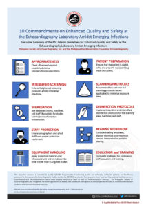
Click here to see infographic, summarising 10 Commandments on Enhanced Quality and Safety at the Echocardiography Laboratory Amidst Emerging Infections.
Guidelines Summaries
PSE Interim Guidelines for Enhanced Quality and Safety at the Echocardiography Laboratory Amidst Emerging Infections
Jose Donato A. Magno, MD, FPSE, FASE;1, 2, 3 Mylene U. Cornel, MD, FPSE;4, 11, 13 Jehan Karen G. Sumalpong, MD, FPSE;5 Emmet Vi Ladlad Pua, MD, FPSE;3 Joyce S. Jumangit, MD, FPSE;3, 6 Rylan Jasper Ubaldo, RMT, FPSE;3 Jonnie Bote Nunez, MD, FPSE;3 Patrick R. Carpo, MD, FPSE;4, 7, 14 Aurora Muriel S. Gamponia, MD, FPSE;3 Viannely Berywn F. Flores, MD, FPSE;3 Aileen C. Senga, RMT, FPSE;3 Celia Catherine C. Uy, MD, FPSE;2, 8 Kristine Hashim Bantala-Supnet, MD, FPSE;3, 9 Karla Rhea G. Rillera-Posadas, MD, FPSE;10 Romeo J. Santos, MD, FPSE;3, 11 Victor L. Lazaro, MD, FPSE;12 Gregorio G. Rogelio, MD, FPSE;4, 13 Stephanie Martha O. Obillos, MD, FPSE;2 Ofelia N. Valencia, MD, FPSE;4, 7 Loewe O. Go, MD, FPSE;13 Benjamin N. Alimurung, MD;14 Rosemarie Ramirez-Ragasa, MD;12 Eugene B. Reyes, MD, FPSE;2, 15 Josephus R. Sibal, MD;1 Jose Jonas D. del Rosario, MD;16 Ma. Bridget D. Fernandez, MD, FPSE;16 Myla Gloria S. Supe, MD, FPSE;4 Edwin S. Tucay, MD, FPSE, FASE 3, 12, 17
1Angeles University Foundation Cardiovascular Institute;
2
Philippine General Hospital;
3
Philippine Heart Center;
4
St. Luke’s Medical Center Global City;
5
Chong Hua Hospital, Cebu City;
6
FEU Nicanor Reyes Medical Foundation;
7
Asian Hospital and Medical Center;
8
ManilaMed – Medical Center Manila;
9
HB Echo Laboratory, Zamboanga City;
10
Notre Dame de Chartres Hospital, Baguio City;
11
Our Lady of Lourdes Hospital, Manila;
12
The Medical City;
13
St. Luke’s Medical Center, Quezon City;
14
Makati Medical Center;
15
Manila Doctors Hospital;
16
Philippine Children’s Medical Center;
17
World Citi Medical Center
10 Commandments on Enhanced Quality and Safety at the Echocardiography Laboratory Amidst Emerging Infections
I. APPROPRIATENESS- All requests for echocardiography must satisfy recognized appropriate use criteria for the adult1, 2 and pediatric3, 4, populations.
- Studies with urgent indications for echo must be prioritized (Appendix A).5, 6 Elective procedures are discouraged and can be rescheduled;5, 7 if such are still highly-indicated, these must undergo strict screening and be aligned with hospital or institutional directives.
- Advanced echo procedures such as transesophageal or stress echocardiography are discouraged due to the higher risk of aerosol generation and must only be performed if crucial in altering clinical management.5, 8
- All requests must first be tagged for suspected or confirmed COVID diagnosis.9 Those with no available confirmatory tests but with alert signs and symptoms suggestive of COVID infection, as well as those with pneumonia or acute respiratory distress syndrome must be strongly flagged.
- Echo labs are advised to enforce clinical pathways or algorithms (Appendix B).4, 8, 10, 11, 12, 13, 14
- Bedside echocardiography is recommended to avoid cross contamination from patient transport.5
- Ideally, machines used for inpatients and outpatients should be different. If there are at least two echo machines, one is ideally dedicated for persons-under-investigation (PUI) and COVID requests. If there is only one machine, rigid disinfection measures must be in place and succeeding echo studies well-spaced out.10 Such dedicated machines should be properly labelled and docked at dedicated and isolated stations.
- If human resource permits, sonographers assigned to handle PUI/COVID requests preferably should not handle non-PUI/COVID requests.10
- The basic minimum protective strategy for all healthcare personnel at the echo lab include: 1) regular hand hygiene (soap and water; 70% alcohol-based rub), 2) proper cough etiquette, 3) medical/surgical face mask, 4) gloves (Appendix C).
- No PPE, no echo study. All sonographers and personnel handling PUI/COVID patients must be in full personal protective equipment, This includes: 1) cap, 2) goggles/face shield. 3) N95 respirator (or higher protection), 4) impermeable full body gown, 5) multi-layer gloves, 6) shoe cover (Appendix C).
- • The echo lab must have a steady supply and inventory system for rational PPE use and have visual aids for proper donning and doffing (Appendix C).14, 15, 16
- Dedicated echo machines (portable units) must be prepared prior to use for PUI/COVID studies.
- Preferably, the entire unit is covered in at least 2 layers of impermeable plastic (waterproof material); inner layer may consist of stretchable plastic (e.g. cling wrap) which can align to contour of unit and not significantly compromise screen view; outer layer may be a 1-piece plastic that covers up to the lower parts of the unit (e.g. possibly up to the wheels).8, 10 (Appendix D)
- Transducer covers can be condoms or commercial transducer covers as long as they fulfill institutionally set infection control guidelines and procedure sterility requirements17
- If the patient is comfortable, a plain surgical/medical facemask should be the bare minimum protection. A mask of higher protective rating is preferred.
- If the patient requires supplemental oxygen, a well-fitting face mask is recommended. For those intubated and mechanically-ventilated, proper ventilator settings must minimize risk of aerosol-generation.
- • Usual standard care is expected, such as adequate thermoregulation using blankets, proper gowning for optimal image acquisition while ensuring patient privacy, and bed railings to prevent falls. Most importantly, the patient should be fairly stable clinically and hemodynamically to be allowed to undergo the focused procedure without interruption.
- Recommend focused over full scanning protocols (particularly for PUI/COVID studies) to minimize exposure time for both patient and sonographer. A study of 5 minutes or less is preferred.
- Be mindful of the specific indication and do targeted study (limited views) to answer the clinical question. The following are the acceptable indications for urgent echo of adults based on international appropriate use criteria: 1) hemodynamic instability or shock (e.g. hypotension, arrhythmia-related instability), 2) acute chest pain syndromes (e.g. myocardial infarction, pulmonary embolism), 3) chest trauma, 4) acute aortic syndromes, 5) acute heart failure (e.g. cardiomyopathy and myocarditis), and 6) acute valve dysfunction (e.g. severe mitral regurgitation, prosthetic valve dysfunction).6 (Appendix A). For patients with COVID, the echo examination should be targeted to assess: 1) regional and global left ventricular function in the setting of acute coronary syndromes or myocarditis, 2) right ventricular size and function especially in suspected acute pulmonary embolism, 3) pericardial effusion amidst inflammatory conditions (myopericarditis), 4) abnormal septal motion or contractility due to new-onset arrhythmias.
- For pediatric patients, only urgent or emergent studies will be performed. The following are some of the other accepted indications for patients with no COVID: 1) cyanotic newborns, 2) transferred patients requiring urgent diagnosis, 3) symptomatic patients seen at the emergency room, 4) pre- or post-surgical patients who require prompt imaging for decision-making.4
- Do not connect ECG cables and instead do time-gated studies. Adjust acquisition time to 3 seconds only.
- The following views are recommended: parasternal window PLAX, PSAX of great vessels; apical window (2-, 3-, 4-, and 5-chamber views) and subcostal window 4-chamber. Added subcostal and suprasternal windows are unnecessary unless they serve as the most optimal windows for acquisition. (Appendix E)
- Consider Spectral Doppler only for very specific indications (e.g. significant stenosis, regurgitation or pulmonary hypertension).
- All measurements must be done offline and not at bedside. Maximize vendor-specific software and technologies to streamline scanning protocol and perform measurements at the workstation.
- All ultrasound machines shall undergo standard disinfection on a daily basis, or intensified disinfection after every use with a patient suspected or confirmed to have COVID-19 infection (opportunities for cleaning/disinfection: at bedside, at the hallway outside the room, and at the docking station.
- The transducer and its cables are most prone to contamination and accumulation of dirt and infective agents. They must be cleaned carefully and diligently using vendor-compatible disinfecting solutions.
- Internal transducers used for invasive procedures (TEE) require routine high-level disinfection, while external transducers used for transthoracic studies require the minimum low-level disinfection.
- Only designated and well-equipped staff (with appropriate PPE) will be allowed to perform the intensified disinfection procedures for the ultrasound machine and the room.
- The reading area/workstation (keyboards, monitors, external devices, chairs, phones, desktops, and door knobs) should be frequently cleaned and disinfected on a daily basis.
- Consider reading templates, digitize workflow, and maximize remote interpretation and data sharing.
- Limit traffic and congestion in the reading room (consider decking schedules). Only the designated echo reader/s (fellow or consultant) for the day should stay in the reading room at any time, taking into consideration proper physical distancing.
- Rapid review and reporting is recommended, with most critical results relayed promptly to the team.
- All healthcare staff must be well-updated regarding such guidelines and well-versed specifically in the use of personal protective equipment.
- Institution-specific protocols and guidelines are recommended, but best practices in close collaboration with other teams and subspecialty societies is likewise encouraged.
- The PSE is committed to uphold quality and safety in echocardiography and shares such a vision with its local and international counterparts.


REFERENCES
1. American College of Cardiology Foundation Appropriate Use Criteria Task Force; American Society of Echocardiography; American Heart Association; ACCF/ASE/AHA/ASNC/HFSA/HRS/SCAI/SCCM/SCCT/SCMR 2011 Appropriate Use Criteria for Echocardiography. A Report of the American College of Cardiology Foundation Appropriate Use Criteria Task Force, American Society of Echocardiography, American Heart Association, American Society of Nuclear Cardiology, Heart Failure Society of America, Heart Rhythm Society, Society for Cardiovascular Angiography and Interventions, Society of Critical Care Medicine, Society of Cardiovascular Computed Tomography, Society for Cardiovascular Magnetic Resonance American College of Chest Physicians. J Am Soc Echocardiogr. 2011;24(3):229–267. doi:10.1016/j.echo.2010.12.008 CrossRef Pubmed
2. Magno JDA. 2018. Clinical Echocardiography Appropriate Use Criteria (The CLEAR Guidelines) for Transthoracic 2D Echocardiography. Section of Echocardiography, Division of Cardiovascular Medicine, Department of Medicine, Philippine General Hospital. Unpublished internal document.
3. Writing Group for Echocardiography in Outpatient Pediatric Cardiology, Campbell RM, Douglas PS, et al. ACC/AAP/AHA/ASE/HRS/SCAI/SCCT/SCMR/SOPE 2014 appropriate use criteria for initial transthoracic echocardiography in outpatient pediatric cardiology: a report of the American College of Cardiology Appropriate Use Criteria Task Force, American Academy of Pediatrics, American Heart Association, American Society of Echocardiography, Heart Rhythm Society, Society for Cardiovascular Angiography and Interventions, Society of Cardiovascular Computed Tomography, Society for Cardiovascular Magnetic Resonance, and Society of Pediatric Echocardiography. J Am Soc Echocardiogr. 2014;27(12):1247–1266. doi:10.1016/j.echo.2014.10.002 CrossRef Pubmed
4. Philippine Heart Center Pediatric Cardiology Non-Invasive Division: Guideline for the Performance of Echocardiography in Pediatric Patients during the COVID-19 Pandemic. March 2020. Unpublished internal document.
5. ASE Statement on Protection of Patients and Echocardiography Service Providers During the 2019 Novel Coronavirus Outbreak. American Society of Echocardiography. April 1, 2020. (https://www.asecho.org/ase-statement-covid-19/)
6. Magno JD, Uy-Agbayani C, Obillos SM, Ragasa R, Anderson B. March 30, 2020. Appropriate use Criteria for Urgent Transthoracic Echocardiography for patients with suspected or confirmed COVID infection (The ACUTE-COVID Echo Guidelines). UP PGH Division of Cardiovascular Medicine, Section of Echocardiography. Unpublished internal document.
7. COVID-19 Clinical Guidance for the Cardiovascular Care Team. American College of Cardiology Clinical Bulletin (S20028-ACCClinical-Bulletin-Coronavirus). March 6, 2020.
8. Philippine Heart Center Division of Noninvasive Cardiology: Guideline for the Performance of Echocardiography during the COVID-19 Pandemic. March 2020. Unpublished internal document.
9. Department of Health. March 16, 2020. Algorithm for triage of patients with possible COVID-19 infection in healthcare facilities. (https://www.doh.gov.ph)
10. Interim Guidance for Intensified Infection Control and Safety Practices at the Echocardiography Laboratory: A Special Policy Document of the Section of Echocardiography, Division of Cardiovascular Medicine, Philippine General Hospital, University of the Philippines Manila. March 2020. Unpublished internal document.
11. Lazaro VL, Bautista AHP. 2020. Guidelines for Echo Technologists in Performing Transthoracic 2D Echocardiography on Confirmed COVID-19, PUI, and PUM Patients. Section of Advanced Echocardiography, Cardiovascular Institute, The Medical City Ortigas. Unpublished internal document.
12. Tucay ES. March 25, 2020. Algorithm in the Screening and Performance of Transthoracic Echocardiography in the Setting of the World Citi Medical Center. Unpublished internal document.
13. FEU-NRMF Medical Center, Section of Cardiology, Department of Medicine. 2020. Guidelines for Cardiac Sonographers doing Echocardiographic Studies on COVID19 Positive Patients/COVID 19 PUI or PUM. Unpublished internal document.
14. Magno JDA et al. March 2020. Guidelines for Heightened Infection Control and Cardiovascular Staff Safety Amidst Emerging Infections: A Special Report of the Task Force for Cardiovascular Quality and Safety of the Angeles University Foundation Cardiovascular Institute. Unpublished internal document.
15. Sequencing for Personal Protective Equipment (PPE). Centers for Disease Control and Prevention; CS250672-E. Available at
16. Liang T and Yu L, editors. (2020). Handbook of COVID-19 Prevention and Treatment. Zehjiang University School of Medicine.
17. American Institute of Ultrasound in Medicine. Guidelines for Cleaning and Preparing External- and Internal-Use Ultrasound Transducers Between Patients, Safe Handling, and Use of Ultrasound Coupling Gel. Nov. 3, 2018. (
ONLINE RESOURCES
1. Philippine Society of Echocardiography:
2. American Society of Echocardiography:
3. Philippine Heart Association: https://philheart.org/index.php/education/pha-covid-resource-center
Copyright Information