
Original Article
October 2026, 35:3
First online: October 2026
Original Article
Initial Experience with Left Bundle Branch Area Pacing in a General Hospital
Supachai Nillugsa,1 Kumpol Chintanavilas, MD 2
Email: rn126@hotmail.com, Telephone: +6682-6692670
2 Corresponding Author, Cardiology Division, Chulabhorn International College of Medicine, Thammasat University, Pathum Thani, Thailand, 12120
Email: kumpol07@tu.ac.th, Telephone: +6662-9265991

ABSTRACT
BACKGROUNDThis study evaluates the procedural success rates, operative learning curves, and post-procedural complications associated with the initial clinical adoption of left bundle branch area pacing (LBBAP) at a provincial cardiovascular tertiary-care center. METHODS
This retrospective cohort study evaluated 56 consecutive patients with clinical indications for permanent pacemaker implantation who underwent left bundle branch area pacing (LBBAP) during the first year of institutional adoption (May 2024 to May 2025) at the tertiary cardiovascular center of Banphaeo General Hospital, Samut Sakhon, Thailand. Primary endpoints included procedural success rates, chronological trends in procedural duration evaluated across four consecutive quarters, and procedure related complications. RESULTS
The cohort (median age 73.5 years; 55.4% female) presented with a high prevalence of non-communicable diseases (82.5%). Main indications were complete heart block (46.4%) and sick sinus syndrome (26.8%). Successful LBBAP capture—evidenced by a left ventricular activation time (LVAT) and a right bundle branch block morphology—was achieved in 85.7% of cases. Electrophysiological outcomes showed a significant reduction in mean QRS duration post-implantation (p < 0.001), with 89.5% of patients achieving a QRS duration < 130 ms. Chronological analysis revealed a clear operative learning curve; median procedural times steadily declined by 32.6%, from 95.0 minutes in Quarter 1 (n=14) to 65.0 minutes in Quarter 4 (n=14), alongside a narrowing of data variability. The overall complication rate was 1.8%, consisting of a single lead dislodgement 10 days post-procedure. CONCLUSION
LBBAP demonstrates high procedural success and a favorable safety profile, with efficiency improving significantly through accumulated operator experience. Keywords
Left bundle branch area pacing; Operative learning curve; Provincial hospital; Pacemaker implantation; Procedural safety.
INTRODUCTION
Historically, conventional dual-chamber permanent pacing has relied on lead fixation at the right ventricular (RV) apex or RV septum. However, cumulative clinical evidence indicates that conventional RV pacing disrupts natural electrical propagation, inducing non-physiological ventricular dyssynchrony that substantially elevates the risk of pacemaker-induced cardiomyopathy (PICM) and subsequent heart failure.1 To circumvent these deleterious long-term sequelae, left bundle branch area pacing (LBBAP) has emerged as a revolutionary physiological pacing alternative. By deploying the pacing lead deeply into the interventricular septum to directly capture the native left bundle branch conduction system, LBBAP achieves a physiological and highly synchronized ventricular activation pattern, thereby preserving left ventricular systolic function.2-4
Since its clinical introduction, the procedural success rate of LBBAP has demonstrated an upward trajectory in large scale literature, rising from approximately 89% in pioneering cohorts to 90%–95% in recent multi-center meta-analyses.5-7 Nevertheless, the vast majority of these robust data are derived from high-volume tertiary academic centers with extensive electrophysiological expertise. Evidence evaluating the clinical translation, procedural feasibility, and safety profiles of initiating an LBBAP program within low-volume, provincial hospitals remains notably scarce. To address this gap in real-world clinical practice, this study aims to evaluate the procedural success, electrocardiographic outcomes, and short-term safety profile during the initial adoption of LBBAP at Banphaeo General Hospital, a lower-volume provincial cardiovascular tertiary-care center utilizing a standard electrocardiogram-guided catheterization laboratory workflow.
METHODS
Study Design and Patient Population
This retrospective cohort study evaluated the initial clinical experience of left bundle branch area pacing (LBBAP) implementation at Banphaeo General Hospital, Samut Sakhon Province. All procedures were performed by an experienced electrophysiologist who was naive to the left bundle branch area pacing (LBBAP) technique. The study population comprised consecutive patients aged ≥ 18 years who possessed standard clinical indications for permanent pacemaker (PPM) implantation—including complete heart block (CHB), sick sinus syndrome (SSS), and high-grade atrioventricular conduction disorders—and underwent LBBAP during the first year of institutional adoption between May 2024 and May 2025.
Procedural Outcomes and Technical Definitions
The primary outcome of interest was the procedural success rate of LBBAP. Successful left bundle branch area capture was strictly defined according to established electrophysiological criteria, requiring a post-procedural 12-lead electrocardiogram (ECG) demonstrating a right bundle branch block (RBBB) pattern in the precordial leads accompanied by a left ventricular activation time (LVAT) of <80 ms. Secondary outcome measures included: The absolute change in QRS duration (ms) from baseline to post-implantation. Total procedural duration (minutes), tracking chronological operational efficiency. The incidence of acute and short-term procedure-related complications.
Statistical Analysis
Statistical analyses were performed using STATA software (version 17.0; StataCorp, College Park, TX, USA). Data distribution normality was verified prior to analysis. Continuous variables were expressed as means ±standard deviations or medians with interquartile ranges where appropriate, while categorical variables were presented as absolute frequencies and percentages. To evaluate the clinical efficacy of pacing on ventricular synchronization, a paired t-test was employed to compare pre- and post-implantation mean QRS durations. To characterize the operative learning curve and proficiency progression, the study cohort was divided into four successive chronological quarters based on the sequential date of the procedure. Because procedural durations exhibited a non-normal distribution, differences in operative times across the four distinct quarters were analyzed using the non-parametric Kruskal-Wallis H test. For all statistical evaluations, a two-tailed p-value of <0.05 was considered statistically significant.
RESULTS
Baseline Demographics and Clinical Characteristics
A total of 56 patients who underwent permanent pacemaker implantation via left bundle branch area pacing (LBBAP) were evaluated. The baseline clinical and demographic characteristics of the study cohort are summarized in Table 1. The cohort demonstrated a slight female predominance (n = 31, 55.4%), with a median age of 73.5 years (interquartile range [IQR], 65.0-79.0 years).
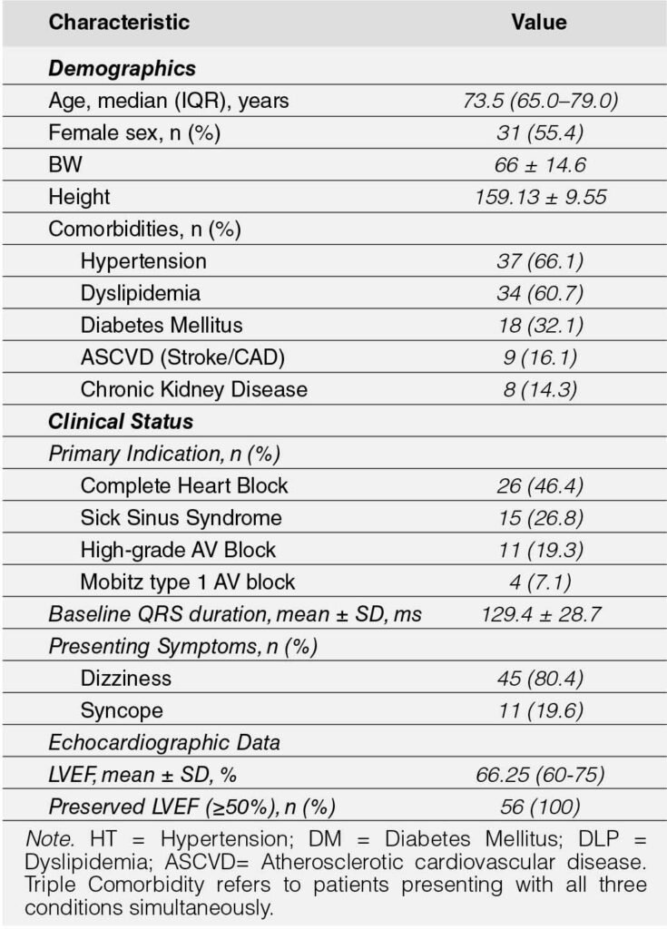
Chronic cardiovascular comorbidities were highly prevalent, affecting 82.5% of the study population. Hypertension was the most frequently observed condition (n = 37, 66.1%), followed by dyslipidemia (n = 34, 60.7%). Notably, a triple comorbidity of concurrent diabetes mellitus, hypertension, and dyslipidemia was present in 21.4% (n = 12) of the patients. Pre-procedural echocardiographic assessment revealed preserved left ventricular (LV) systolic function (LVEF≥50%) in all of the patients, with a mean overall cohort left ventricular ejection fraction (LVEF) of 66.25%.
Clinical Indications and Presentations
The primary clinical indications necessitating permanent cardiac pacing were complete heart block (46.4%), followed by sick sinus syndrome (26.8%), and high-grade atrioventricular (AV) block (19.3%). Prior to the intervention, the most common symptomatic presentation driving clinical evaluation was dizziness, which was reported by 73.7% of the patient population.
Procedural and Electrophysiological Outcomes
Successful LBBAP capture was confirmed by a Left Ventricular Activation Time (LVAT) of <80 ms with ICBBB defined by the V6–V1 interval 33-55 ms in 85.7% of patients, indicating successful left-to-right ventricular conduction. The mean QRS duration significantly decreased from 129.6 ±3.15 ms at baseline to 113.5±3.15 ms following the procedure (mean difference: 16.05 ms; 95% CI, 7.21 to 24.88; p = .0001). As shown in figure 1. Approximately 91.1% (51 patients) achieved a post-procedural QRS duration of ≤130 ms.
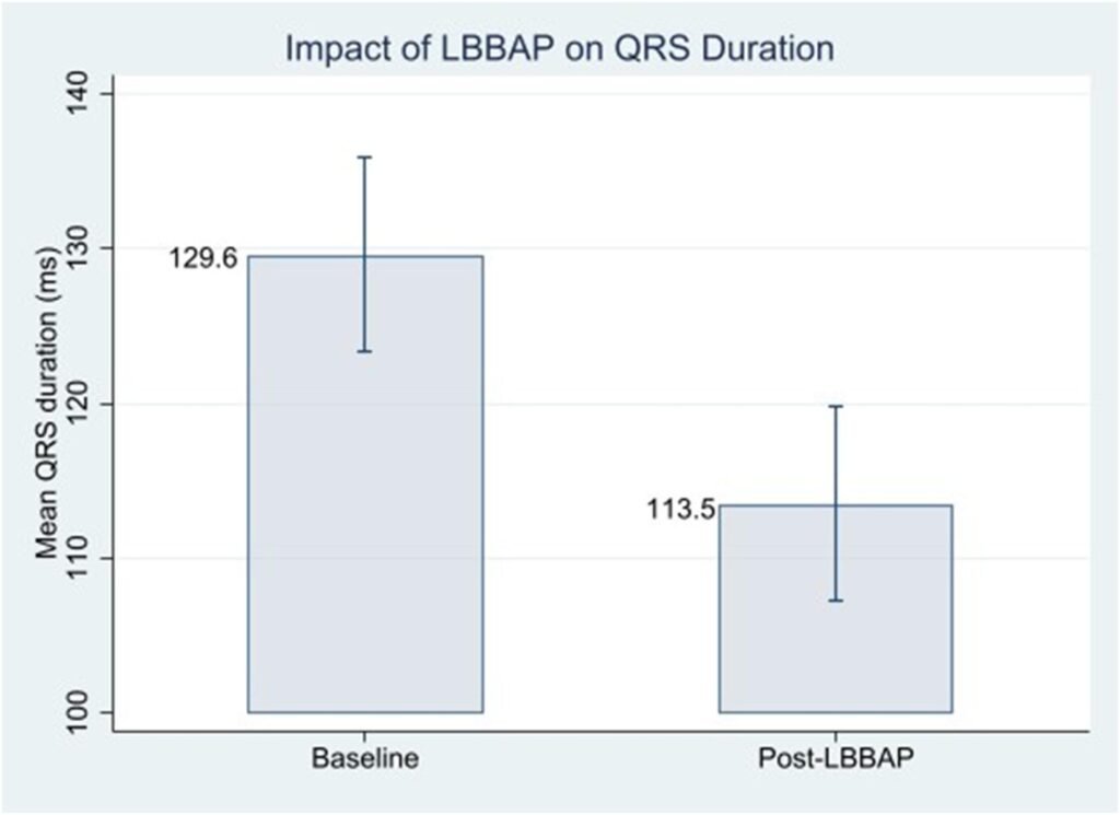
Error bars represent 95% Confidence Intervals. ***p < 0.001 vs. baseline.
Learning Curve and Operational Efficiency Analysis
A chronological evaluation of the initial implementation phase demonstrated a progressive and steady downward trend in total procedural durations over the study period (Figure 2). During the first chronological quarter (n = 14), the median procedural time was 95.0 minutes. This value decreased incrementally to 77.5 minutes in the second quarter (n = 14), 72.5 minutes in the third quarter (n = 14), and reached a minimum median of 65.0 minutes by the final quarter (n = 14). Although a numerical reduction in medians was apparent, a Kruskal-Wallis H test indicated that the overall difference in procedural times across the four distinct quarters did not reach statistical significance (χ2(3) = 6.96, p = .073). This distribution was heavily influenced by a wider dispersion of operative durations and distinct upperbound outliers exceeding 140 minutes in the final quarter, reflecting procedural variability inherent to the late initiation phase (Figure 2).
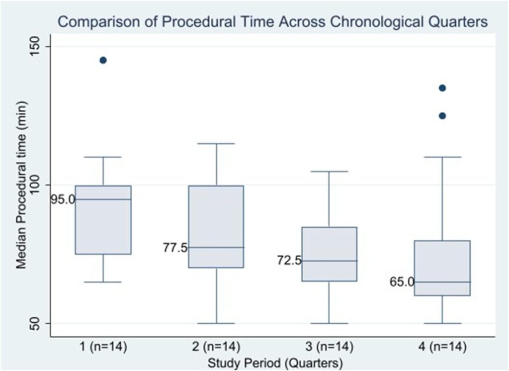
Box-and-whisker plots illustrate the median procedural duration (minutes), interquartile ranges (IQRs), and outlying data points across the four successive quarters of the initial study period.
Concurrently, analysis of fluoroscopic exposure times across the four chronological quarters mirrored this downward trajectory. The median fluoroscopic duration decreased from 8.2 minutes (interquartile range [IQR]: 5.5–13.5 min) in the first quarter (n = 14) to a minimum of 4.2 minutes (IQR: 3.2–6.9 min) by the final quarter (n = 14). Similar to the overall procedural times, this chronological reduction did not reach statistical significance via the Kruskal–Wallis H test (χ2(3) = 7.12, p = .068). This lack of significance was primarily driven by prominent upper-bound outliers during the fourth quarter—most notably a single case reaching 46.2 minutes which expanded the total variance despite a consistently narrowing interquartile range among standard cases (Figure 3).
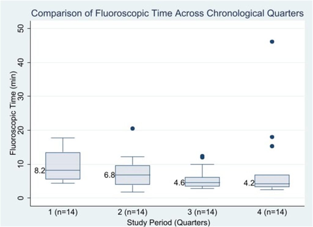
Box-and-whisker plots depict the median fluoroscopic exposure times (minutes), IQRs, and extreme statistical outliers categorized by chronological procedural sequence.
Safety and Complications
The clinical safety and complication profile of the initial adoption of left bundle branch area pacing (LBBAP) at Banphaeo General Hospital was highly favorable. No acute or major complications occurred perioperatively, within the first 24 hours, or up to 30 days post-procedure. Across the entire evaluated cohort, the sole recorded adverse event was a single case of late lead dislodgement (1.8%) occurring 10 days post-implantation. This incident was successfully managed via standard percutaneous lead repositioning without further adverse sequelae or long-term clinical compromise.
DISCUSSION
This retrospective study evaluates the clinical translation, procedural feasibility, and safety profiles of initial institutional adoption a left bundle branch area pacing (LBBAP) within a lower-volume provincial cardiovascular tertiary-care center utilizing a standard electrocardiogram-guided catheterization laboratory workflow. Our principal findings demonstrate that LBBAP initiation is highly feasible and safe, achieving a high procedural success rate of 85.7% and an exceptionally low short-term complication rate (1.8%).
The 85.7% procedural success rate observed during our first-year implementation phase aligns closely with pioneering multi-center cohorts and large-scale registries, which historically report success rates ranging between 89% and 95%. (5-7) Crucially, the vast majority of existing LBBAP data originate from high volume tertiary academic medical centers with extensive electrophysiological resources and specialized operator experience. However, our real-world data demonstrate that a lower volume provincial hospital can replicate these rigorous electrophysiological metrics. This success underscores the robustness and reproducibility of standard ECG-guided implantation techniques, proving that extensive subspecialty volume is not a mandatory prerequisite to establishing a safe and clinically effective conduction system pacing program.
From an electrophysiological perspective, the primary therapeutic objective of LBBAP is to circumvent the non-physiological ventricular dyssynchrony and subsequent pacemakerinduced cardiomyopathy (PICM) frequently caused by traditional right ventricular apex pacing. In our cohort, this physiological synchronization was objectively confirmed by a significant reduction in mean QRS duration from 129.6 ± 3.15 ms at baseline to 113.5±3.15 ms post-implantation (p = .0001). By directly capturing the native conduction system, as evidenced by a left ventricular activation time (LVAT) of <80 ms and characteristic precordial incomplete right bundle branch block (ICBBB) patterns, the technique preserves or restores physiological left-to-right ventricular propagation. 91.1% of patients achieving an optimal QRS duration of ≤ 130 ms.
A key contribution of this study is the granular, chronological characterization of the operative learning curve during the program’s initiation phase. We observed a distinct and continuous numerical decline in median procedural durations, decreasing from 95.0 minutes in the first quarter to 65.0 minutes by the final quarter, representing a 32.6% improvement in procedural efficiency. Interestingly, while a clear downward trend in medians was apparent for both operative and fluoroscopic times, non-parametric statistical evaluation did not yield statistical significance across the quarters (p = .073 for procedural time; p = .068 for fluoroscopic time). Our findings highlight a critical statistical paradox that frequently characterizes the clinical adoption of novel cardiac interventions. While the overall analysis did not achieve statistical significance due to prominent upper-bound outliers in the final quarter, these data must be interpreted through a clinical lens. Rather than reflecting a lack of technical proficiency, these late-stage outliers represent a standard evolutionary phase in the operator’s learning curve: as routine cases are mastered in Quarters 1–3, operator confidence increases, leading to the deliberate selection of highly complex anatomical variations and challenging substrates in Quarter 4. This interpretation is firmly supported by the steady narrowing of the IQRs across successive quarters, which confirms that procedural efficiency was optimizing and stabilizing for the vast majority of standard cases.
The clinical safety profile demonstrated during our first-year experience further supports the safety of migrating LBBAP to a lower-volume provincial cardiovascular tertiary-care center utilizing a standard electrocardiogram-guided catheterization laboratory workflow. No acute, major perioperative complications, such as acute septal perforation, coronary artery injury, or cardiac tamponade, were encountered. The sole adverse event recorded was an isolated case of late lead dislodgement (1.8%) occurring 10 days post-procedure, which was successfully managed via standard percutaneous repositioning without long-term sequelae. This remarkably low complication rate satisfies international safety benchmarks and confirms that the technical demands of deep septal lead deployment do not inherently compromise patient safety when introduced systematically into a new center.
Study Limitations
This study has several limitations that should be acknowledged. First, it was a single-center, retrospective cohort study with a relatively modest sample size (N = 56), which reflects the initial year of institutional adoption in a lower-volume provincial cardiovascular tertiary-care center. Second, the follow-up window was restricted to the short-term post-procedural period (30 days), preventing the evaluation of long-term lead performance parameters (such as chronic capture thresholds and sensing stability) or hard clinical endpoints (including heart failure hospitalizations and all-cause mortality). Finally, because this study lacked a contemporary parallel control group undergoing conventional right ventricular pacing, direct comparative assertions regarding long-term clinical superiority within this specific provincial setting cannot be definitively made. Future prospective, multi-center trials with extended follow-up durations are warranted to confirm the long-term clinical and economic benefits of widespread LBBAP adoption in community healthcare infrastructures.
CONCLUSION
The initiation of an LBBAP program in a lower-volume provincial cardiovascular tertiary-care center utilizing a standard electrocardiogram-guided catheterization laboratory workflow is safe, feasible, and clinically effective. The technique achieves high procedural success rates, favorable post-implantation electrical synchronization, and minimal complications. Procedural efficiency steadily optimizes within the first year, establishing LBBAP as a viable first-line physiological pacing modality outside of specialized tertiary academic centers.
REFERENCES
1. Boyle TA, Pothineni NVK, Austin M, et al. Incidence and predictors of pacing-induced right ventricular cardiomyopathy. Circ Arrhythm Electrophysiol. 2024;17(10):e013070. doi:10.1161/CIRCEP.124.013070 CrossRef Pubmed
2. Yin L, Wang L, Meng J, et al. A systematic review and meta-analysis of the impact of left bundle branch area pacing on right ventricular function. Front Cardiovasc Med. 2025;12:1545757. Published 2025 Jan 30. doi:10.3389/fcvm.2025.1545757 CrossRef Pubmed
3. Vijayaraman P, Cano O, Ponnusamy SS, et al. Left bundle branch area pacing in patients with heart failure and right bundle branch block: Results from International LBBAP Collaborative-Study Group. Heart Rhythm O2. 2022;3(4):358-367. Published 2022 May 14. doi:10.1016/j.hroo.2022.05.004 CrossRef Pubmed
4. Yu GI, Kim TH, Cho YH, et al. Left bundle branch area pacing in mildly reduced heart failure: A systematic literature review and meta-analysis. Clin Cardiol. 2023;46(7):713-720. doi:10.1002/clc.24028 CrossRef Pubmed
5. Padala SK, Master VM, Terricabras M, et al. Initial Experience, Safety, and Feasibility of Left Bundle Branch Area Pacing: A Multicenter Prospective Study. JACC Clin Electrophysiol. 2020;6(14):1773-1782. doi:10.1016/j.jacep.2020.07.004 CrossRef Pubmed
6. Parlavecchio A, Vetta G, Coluccia G, et al. Success and complication rates of conduction system pacing: a meta-analytical observational comparison of left bundle branch area pacing and His bundle pacing. J Interv Card Electrophysiol. 2024;67(4):719-729. doi:10.1007/s10840-023-01626-5 CrossRef Pubmed
7. Ellenbogen KA, Fagan DH, Zimmerman P, Vijayaraman P. Left bundle branch area pacing using a lumenless lead: a systematic literature review and meta-analysis. J Cardiovasc Electrophysiol. 2024;35:1721-1735. doi:10.1111/jce.16287 CrossRef Pubmed
Copyright Information