Case Report
Dec 2022, 31:2
First online: 31 December 2022
Case Report
Single Coronary Anomaly detected by Coronary Computed Tomography Angiography in an Elderly Female with Angina
Rainier John S. Buensalida,1 Neil Pecache,1 Kristina Marie Michelle O. Rivera,1 Victor Lazaro,1 Simonette Kristine T. Sawit1
Main author: Rainier John S. Buensalida
Email address: rjbuensalidamd@outlook.com
ABSTRACT
A 76-year-old female was admitted for new onset angina and diagnosed with a rare single coronary anomaly detected by coronary computed tomography angiography. Coronary computed tomography angiography also revealed mild coronary atherosclerosis with no significant stenoses. The patient was managed conservatively with beta blocker, ACE inhibitor and statins. She was discharged improved and remains asymptomatic on follow up.Keywords
coronary artery anomaly; single coronary artery; CCTA
INTRODUCTION
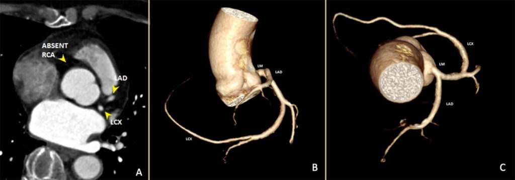
A 76-year-old female, known hypertensive, was at the emergency room with chest pain on exertion and palpitations. Physical examination was unremarkable. Electrocardiogram showed normal sinus rhythm and non-specific ST-segment changes. Serial high-sensitivity troponin were normal. Echocardiogram showed normal left ventricular size and systolic function with no regional wall motion abnormalities. On coronary computed angiography (CCTA) there was a single coronary artery (SCA) arising from the left coronary sinus of valsalva that bifurcated into a type III left anterior descending artery and a moderate-sized left circumflex artery that extended and traversed the base of the heart, then terminated at the right atrioventricular groove to supply marginal branches to the right ventricle (Figure 1). This was consistent with a Type L-I pattern based on the modified Lipton’s classification for isolated SCA.1 CCTA also revealed mild coronary atherosclerosis with no significant stenoses. The patient was managed conservatively with beta blocker, ACE inhibitor and statins. She was discharged improved and remains asymptomatic on follow up.
DISCUSSION
This case demonstrated the incidental finding of SCA arising from the left sinus of valsava, a rare congenital anomaly found in less than 1% of CCTA and conventional coronary angiography studies.1-4 SCA may be asymptomatic at the time of diagnosis or have variable presentation, ranging from mild non-specific symptoms up to sudden cardiac death, especially during exercise.4 About 15% of isolated SCA may have myocardial ischemia directly caused by the abnormal anatomy of the arteries and not necessarily by coronary artery disease.2
Coronary atherosclerosis is generally uncommon in younger SCA patients, but accelerated atherosclerosis may occur in older patients, resulting from turbulent blood flow and predisposition to endothelial injury.5
Prognosis for patients with SCA varies from excellent with no decrease in life expectancy to sudden death.3-5 Unlike other classifications where coronary perfusion might be compromised, the R-I or L-I classification types (solitary vessel arising from either left or right coronary cusp, following the course of either a normal right or left coronary artery) usually have a benign clinical course.2-5
In conclusion, in the absence of significant coronary artery disease and other congenital anomalies, this elderly female with an incidental finding of isolated SCA L-I type had a benign clinical course on conservative medical management. To our knowledge, this is the first local case report of SCA detected by CCTA in an elderly female with angina.
REFERENCES
1. Yamanaka O, Hobbs R, Coronary artery anomalies in 126,595 patients undergoing coronary arteriography, Catheterization and Cardiovascular Diagnosis, 21(1):28-40, 1990. CrossRef Pubmed
2. Umairi R, Al-khouri M, Prevalence, Spectrum, and Outcomes of Single Coronary Artery Detected on Coronary Computed Tomography Angiography (CCTA), Hindawi Radiology and Research, https://doi.org/1o.1155/2019/2940148, 2019. CrossRef
3. Elbadawi A, Baig B, Elgendy I, Alotaki E, Mohamed A, Barssoum K, Fries D, Khan M, Khouzam R, Single Coronary Artery Anomaly: A Case Report and Review of Literature, Cardiology and Therapy, 7:119–123, 2018. CrossRef Pubmed
4. Arslan U, Karamanlıoğlu M, Korkmaz A, Conventional and computed tomography angiography views of a rare type of single coronary artery anomaly: right coronary artery arising from distal left circumflex artery, Anadolu Kardiyol Derg, 12: 517-524, 2012.
5. Chou P, Kao C, Lee M, Lin S, Right Coronary Artery Originating From Distal Left Circumflex Artery in a Patient With an Unusual Type of Isolated Single Coronary Artery, Japan Heart Journal 45(2):337-42, 2004 CrossRef Pubmed
Copyright Information